The Radiological Research Trust was registered with the Charity Commission (registration number 292828) in October 1985. The Trust was set up to raise funds and distribute grants for research and education in Medical Imaging, in response to the severe lack of funding for such a key medical field as radiology. The RRT has now (February 2025) decided to merge its funds with the British Institute of Radiology to form the George du Boulay Pump-Priming Award. (see statement below). The BIR will administer this fund.

Statement:
The Radiological Research Trust (RRT) is pleased to announce that it will be transferring its funds to the British Institute of Radiology (BIR) supporting the newly created BIR George du Boulay Pump-Priming Award. This transition ensures the continuation of our commitment to supporting innovative radiological research, facilitating vital early-stage funding to advance cutting-edge projects in the field.
Professor George du Boulay CBE, FRCR, FRCP, was a pioneering academic neuroradiologist and a founding member of the British Society of Neuroradiologists. He served as Professor of Neuroradiology at the Institute of Neurology and Head of the Lysholm Department of Radiology at the National Hospital for Neurology and Neurosurgery from 1975 until 1984. In 1985, he established the RRT to address the severe lack of funding for research and education in medical imaging, becoming its first director.
Over the years, the RRT has supported numerous pioneering projects, including:
- Magnetic Resonance Imaging (MRI) Bone Mineral Density: Technical validation of a novel MRI technique for quantifying bone mineral density, which has potential clinical utility for disorders causing both new bone formation and bone destruction.
- Juvenile Idiopathic Arthritis: Development of quantitative MRI methods to assess this condition, aiding in better diagnosis and treatment planning.
- Prostate Haemorrhage: Investigation into post-biopsy prostate haemorrhage and its effect on prostate MRI, emphasizing the importance of MRI scan analysis before and after prostate biopsy.
- Whole-Body MRI for Paediatric Lymphoma: Research into whole-body MRI imaging for staging paediatric lymphoma, demonstrating that whole-body MRI is a viable alternative to imaging techniques using ionising radiation.
- COVID-19 Stroke Apical Lung Examination Study: A national prospective study analysing lung apices in patients presenting with suspected acute stroke, contributing to better management of patients with an acute stroke during the pandemic.
These projects have led to significant advancements in medical imaging, including international recognition, large-scale funding from organizations such as Cancer Research UK and The Wellcome Trust, and numerous publications.
We are confident that the BIR will carry forward this legacy with the same dedication and vision, continuing to support groundbreaking research in radiology.
Please check the BIR website for details as this is established. https://www.bir.org.uk
RRT Trustee Professor Margaret Hall-Craggs – President of the International Society of Magnetic Resonance in Medicine
See below for some grant holder feedback of the value to individuals of RRT funded projects, including some of the difficulties caused by the Covid pandemic.
Dr T. Ratneswaren (2024). COVID-19 Stroke Apical Lung Examination Study 2: a national prospective CTA biomarker study of the lung apices, in patients presenting with suspected acute stroke (COVID SALES 2)
‘The COVID-SALES 2 study is the follow-up to the landmark first COVID-SALES study (published in 2020). Undertaken in collaboration with the Radiology Academic Network for Trainees (RADIANT), this prospective multicentre study expanded on the scope of the first study, with data collected from 13 UK hyperacute stroke units. The findings have reinforced the clinical utility of lung apical changes on routine CT angiography studies in the incidental diagnosis of COVID-19.’ Jason Mak, Clinical Radiology SpR, RADIANT Co-chair.
‘In summary, we recommend routinely analysing lung apices in patients with stroke undergoing carotid CTA, because this rapid and easy assessment of apices is valuable, opportunistic, and “free” information in a routine and unmodified scan. The apical analysis simply identifies patients likely to have COVID-19 with early downstream benefits such as limiting disease transmission.’ Dr Thomas Booth, Reader in Neuroimaging, School of Biomedical Engineering & Imaging Sciences
https://www.sciencedirect.com/science/article/pii/S2213158224000299?via%3Dihub
This study was partly funded by RRT
Dr. Nathan Chan (2020): ‘COVID-19 Stroke Apical Lung Examination prospective validation Study: A Diagnostic and Prognostic Imaging Biomarker in Suspected Acute Stroke (COVID SALES)’
On behalf of the Radiology Academic Network for Trainees (RADIANT) and the COVID-19 stroke apical lung examination study (COVID SALES) team including Dr Tom Booth, Dr Tarini Ratneswaren and Mr John Aeron-Thomas, I would like to thank the Radiological Research Trust (RRT) for their most generous contribution to our work. Apical lung changes on CT angiography suggestive of COVID-19, have been shown to be a promising diagnostic and prognostic biomarker. The aim of this study was to test this in different centres across the UK. The biomarker is free information obtained opportunistically from a routine urgent stroke scan. It is available very early on in the patient pathway well before SARS COV-2 PCR test results are ready and will contribute to better management of acute stroke patients.
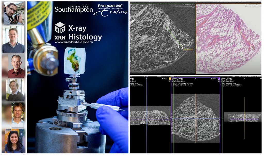
Dr. Orestis Katsamenis (2020): ‘Pathogenesis of lethal respiratory COVID-19; 3D X-ray histology imaging of pathology wax blocks’
I am beamline scientist and a clinical applications lead at the μ-VIS X-ray Imaging Centre (www.muvis.org) at the University of Southamtpon running the 3D X-ray Histology facility. In 2020 I was awarded a seed funding from the Radiological Research Trust (RRT) to study the pathogenesis of lethal respiratory COVID-19 by means of a novel high-resolution X-ray CT imaging modality that allows non-destructive 3D imaging of tissue biopsies. The technique, a.k.a 3D X-ray Histology (www.xrayhistology.org) was developed at University of Southamtpon and I have been working on its development since c. 2015. The RRT funding was pivotal as it allowed us to overcome the primary cost-related obstacles associated with sample preparation, imaging cost, international shipments, and other consumables and allowed me to bring together a multidisciplinary team of experts from the University of Southamtpon, UK and Erasmus University Medical Center, NL, and kick-start the project. Together, we study the pathogenesis of micro-thrombotic events caused by the SARS-CoV-2 infection in the lungs and develop semi-automated AI-based workflow that allows crosstalk of conventional 2D histology and 3D X-ray Histology, shining new light to the damage caused by COVID-19. Importantly, with appropriate optimisation, the same workflow can be applied for the study and/or characterisation of other pathologies beyond COVID-19. RRT support has been vital in allowing me to establish a long-lasting international collaboration, diffuse UK-developed technology to European partners and equally “bring in” expertise from one of the best Pathology centres in EU.
The team: O.L. Katsamenis1, P. Lackie1, P. Schneider1, J. von der Thüsen2, P. Ciet2, E. Ho1,4 & T.E. Trandafir2, B. Lopuhaä2, F. Akram2, M.J. Lawson4, J.L. Wolf2, E. Konstantinopoulou1, A.P. Stubbs2, Y. Li.2
1University of Southampton, UK; 2Erasmus Medical Center, Rotterdam, NL; 3Rosalind Franklin Institute, Harwell Campus, Didcot, UK; 4University of Manchester, Manchester, UK
The project: Pathogenesis of lethal respiratory COVID-19; 3D X-ray histology imaging of pathology wax blocks
Dr Orestis L. Katsamenis Biomedical Imaging portfolio | Clinical Applications lead
μ-VIS X-ray Imaging Centre | 3D X-ray Histology facility, University of Southampton
Links: www.xrayhistology.org | www.muvis.org | https://www.erasmusmc.nl/en/research/departments/pathology
Tweeter material:
@RRTrust support has been vital in allowing me to establish a long-lasting international collaboration (@unisouthampton U @erasmusmcintl), diffuse UK-developed technology (xrayhistology.org) to EU partners & “bring in” expertise from one of the best Pathology centres in EU.
Dr. Haris Shuaib (2019): ‘Improving treatment of glioblastoma: differentiating progression from pseudo progression’
The grant I received from the Radiological Research Trust was pivotal in launching my PhD work, which has grown to be the largest prospective trial applying machine learning to high grade brain tumours in the UK. The funding allowed me to purchase a GPU-powered machine that was used to develop a machine learning algorithm that can differentiate progression from pseudo-progression in glioblastoma patients. This, alongside the eligibility for CRN support through NIHR portfolio adoption, allowed me to accelerate towards a prospective trial, which is now also supported by an NIHR Doctoral Research Fellowship.
Naomi Sakai (2018): ‘Technical validation of a novel MRI technique for quantificaiton of bone mineral density’
I received a grant from the RRT in 2018 for technical validation of a new MRI technique to measure bone mineral density whilst I was doing a PhD during time out of my clinical radiology training. The grant enabled me to start a small project, developing and scanning a new imaging phantom and scanning volunteer patients. This work was eventually included in my PhD thesis (even though the idea didn’t exist at the start of my PhD, which just demonstrates how PhD theses can evolve) and I was able to present it at an international imaging meeting (ISMRM 2019, Montreal). I am currently taking another break from my clinical radiology training (on maternity leave) and will be taking up an academic clinical lecturer post on my return. The grant I received undoubtedly helped me progress in my academic career, giving me experience of applying for funding and also enabling me to bring an initial idea to reality. I would encourage researchers with an idea to apply as both the application process and ability move the project forward have been beneficial.
Dr. Rosalind Pratt (2017): ‘Advanced MRI for the assessment of placental function in Fetal Growth Restriction’
My research aims to discover whether a safe medical imaging technology (Magnetic Resonance Imaging (MRI)) can be developed to assess how well the placenta is working during pregnancy.Placental insufficiency is a condition where the placenta cannot meet the oxygen and nutritional demands of a growing baby in the womb. This is responsible for over a third of stillbirths in the UK. It is also associated with long term health problems for the baby, including cerebral palsy due to brain injury from low oxygen levels. The mother can suffer from high blood pressure and a life-threatening condition called pre-eclampsia.The research grant I received from RRT helped fund the development of a computational MRI model of placental function that estimates the level of oxygen in the baby’s blood. Using this model we investigated blood oxygen levels in pregnancies with small babies compared to normally grown babies, and found they were significantly lower. I am now an NIHR funded Clinical Lecturer in Fetal Medicine at UCL/UCLH, where I am continuing this important research. I have several grants that are funding a larger study to further investigate the use of MRI to assess placental function. The long-term aim of this research is to establish an effective MRI test of placental function for routine use in clinical practice.
Current Funding
- Wellbeing of Women Sir Victor & Lady Blank Postdoctoral Research Fellowship
- The Academy of Medical Sciences Starter Grants for Clinical Lecturers
- The EGA Hospital Charity
Relevant Papers
- Melboure A*, Aughwane R*, Sokolska M, Owen D, Kendall G, Flouri D, Bainbridge A, Atkinson D, Deprest J, Vercauteren T, David AL, Ourselin S. Separating fetal and maternal placenta circulations using multiparametric MRI. Magn Reson Med, 2019;81:350-61.
- Aughwane R, Mufti N, Flouri D, Maksym K, Spencer R, Sokolska M, Kendall G, Atkinson D, Bainbridge A, Deprest J, Vercauteren T, Ourselin S, David AL, Melbourne A. Magnetic resonance imaging measurement of placental perfusion and oxygen saturation in early-onset fetal growth restriction. BJOG: Int J Obstet Gy2020; doi 10.1111/1471-0528.16387
Tweetable
MRI reveals differences in feto-placental oxygen saturation between normal and FGR pregnancy that is associated with disease severity
Dr. Manil Chouhan (2016): ‘SUPER-RESOLUTION FOR MAGNETIC RESONANCE CHOLANGIO-PANCREATOGRAPHY’
Seed funding from the RRT was instrumental in supporting my early career research, providing me with the opportunity to explore new independently generated research ideas and concepts. The application of super-resolution reconstruction to MRCP yielded one of the most downloaded papers from Magnetic Resonance in Medicine in 2019 and has allowed me to secure substantial further funding from CRUK for ongoing research evaluating the clinical application of this technology to pancreatic cancer.
News Update: We welcome Dr John Ioannou on becoming a new Trustee of the Radiological Research Trust
See the Contact Us page for more information on Dr Ioannou’s background and experience which will help the RRT progress
The Radiology Academic Network for Trainees
** Breaking News: A second Covid research grant has just been awarded to a group from Kings College London **
A special grant for Covid research has been awarded to an international team led by the University of Southampton
https://www.southampton.ac.uk/muvis/media-activities/2021-okatsamenis-rrt-covid.page
Project title: Pathogenesis of lethal respiratory COVID-19; 3D X-ray histology imaging of pathology wax blocks
The team (photo order): Orestis Katsamenis (UoS), Jan von der Thüsen (EMC), Philipp Schneider (UoS), Peter Lackie (UoS), Pierluigi Ciet (EMC), Elaine Ho (UoS)
Institutions: University of Southampton, UK and the Erasmus University Medical Centre, NL
To apply for a Research Grant, please see the Grants page for instructions and use the following document:
RRT Research Grant Application Form
Below are comments on the RRT from a couple of recent early career researcher awardees. See the Research Page for more details and other previous awards.
“This is an excellent funding opportunity to start a new project”
Naomi Sakai, University College London
Technical validation of a novel MRI technique for quantification of bone mineral density

presenting results from their RRT funded projects at an imaging conference
“RRT is a great place to apply for your first funding and can be a springboard for future grants.”
Tim Bray, University College London
Quantitative magnetic resonance imaging in juvenile idiopathic arthritis

showing Oedema and Fat Metaplasia by measuring Fat Fraction and Relaxation Time R2*
What is Radiology?
Radiology is a specialty of medicine that just about everybody in their life uses at some point. If you have ever broken a bone, had a baby, found a lump that has been investigated for cancerous cells or your child as swallowed something horrible you will have used radiological related equipment and expertise.
Radiology has been used for medical purposes for over a century and remains a cornerstone of medicine. It can be used for not only diagnostic but also therapeutic purposes. Physicians use the information gained through radiological imaging to diagnose, treat and monitor injuries and diseases.
Radiology is critical to advances in medicine and therefore ultimately the care which hospitals can provide you, their patients. Radiologists use a wide range of methods to study the body including Ultrasound (US), X-Ray Computed Tomography (|CT) and Magnetic Resonance Imaging (MRI) as seen in the images on the right.
Today the aims of the Trust remain steadfast in providing critical funding to such an important specialty of medicine. Demand for funding remains high and the Trust remains committed to supporting some of the most exciting research in the field.

Donate to the Radiological Research Trust

![]()